
Preclinical, Clinical, and Translational Sciences
 photo")
Bharatbhai N. Chaudhary, M.Pharm. (he/him/his)
Graduate Student
University of Nebraska Medical Center
Omaha, Nebraska, United States
Bharatbhai N. Chaudhary, M.Pharm. (he/him/his)
Graduate Student
University of Nebraska Medical Center
Omaha, Nebraska, United States
Sudipta Panja, Ph.D.
Assistant professor
University of Nebraska Medical Center
Omaha, Nebraska, United States
 photo")
Mohammad Uzair Ali, B.Pharm. (he/him/his)
Graduate Student
University of Nebraska Medical Center
Omaha, Nebraska, United States
Soumya S. Dey, MS
Graduate Student
University of Nebraska Medical Centre
Omaha, Nebraska, United States
Howard Gendelman, M.D.
Professor
University of Nebraska Medical Center
Omaha, Nebraska, United States
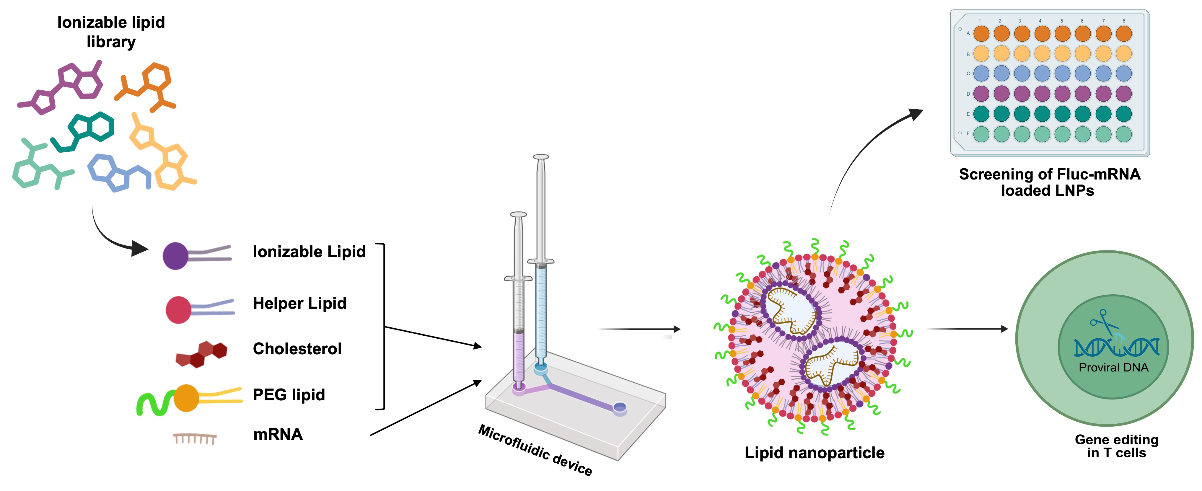 Figure 1. Schematic representation of LNP formulation using a microfluidic device. The illustration also depicts the preliminary screening of Fluc-mRNA-loaded LNPs and the gene delivery of CRISPR-Cas9-encapsulated LNPs into T cells.
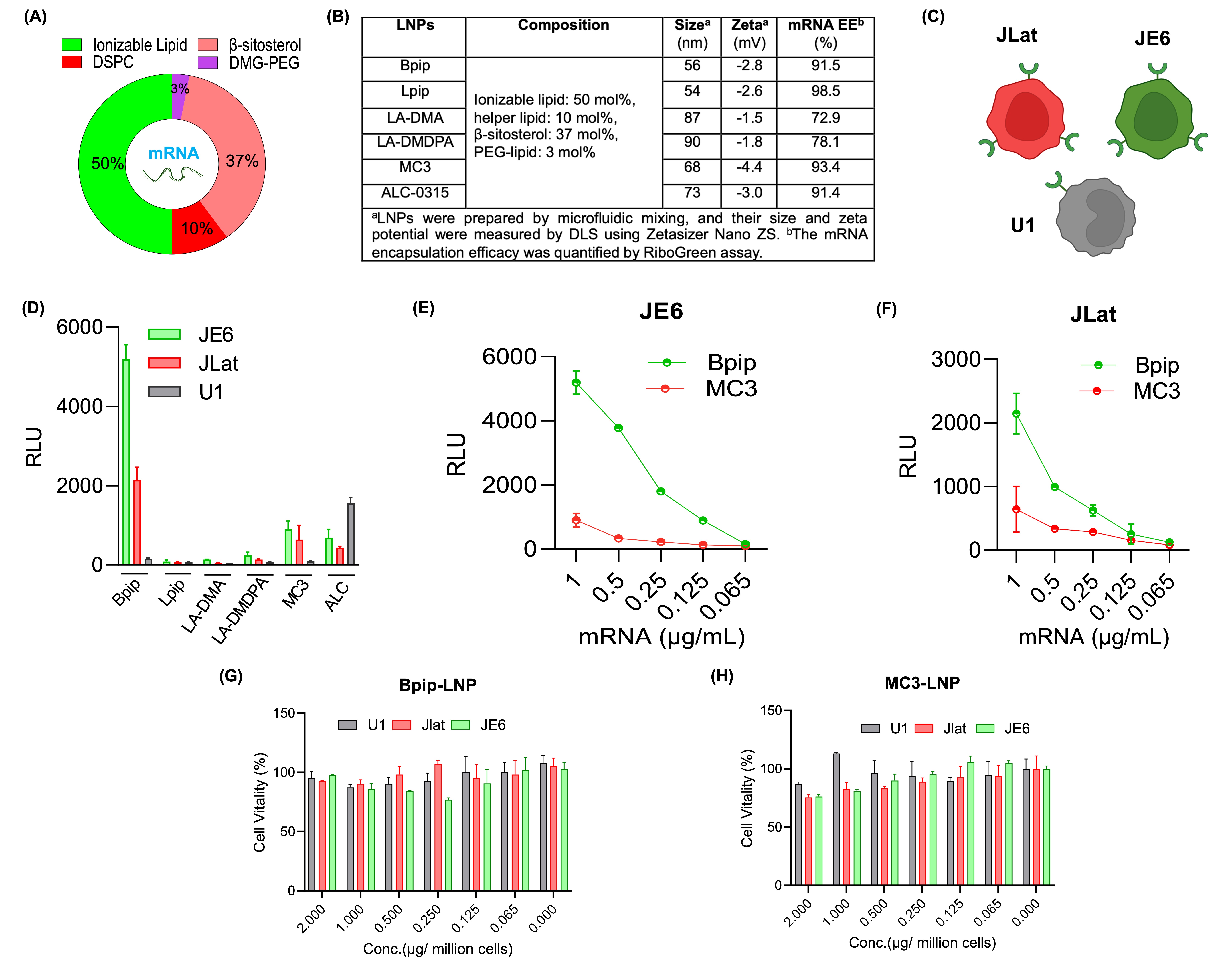 Figure 2. Preparation and preliminary screening of LNPs in vitro. (A) Donut charts showing the molar composition of the lipids used to formulate LNPs. (B) Size, PDI, and zeta potential of LNP measured by dynamic light scattering (DLS) and mRNA encapsulation efficiency measured by RiboGreen assay. (C) Schematic representation of latent HIV-1-infected cells (T-lymphocytic JLat, JE6, and promonocytic U1). (D) Luciferase mRNA translation efficacy of LNPs in the JE6, JLat, and U1 cells. The mRNA translation efficiency was assessed 48 hours post-LNP treatment (at 1µg/ million cells mRNA dose) using the luciferase assay and presented as relative luminescence units (RLU). (E, F) Dose-dependent mRNA translation efficiency of Bpip and MC3 LNPs in JE6 and JLat cells. (G, H) The mRNA dose-dependent cell vitality of Bpip and MC3 LNPs in U1, JLat, and JE6 cells. LNPs show more than 80% cell vitality at a dose of 1µg/106 cells.
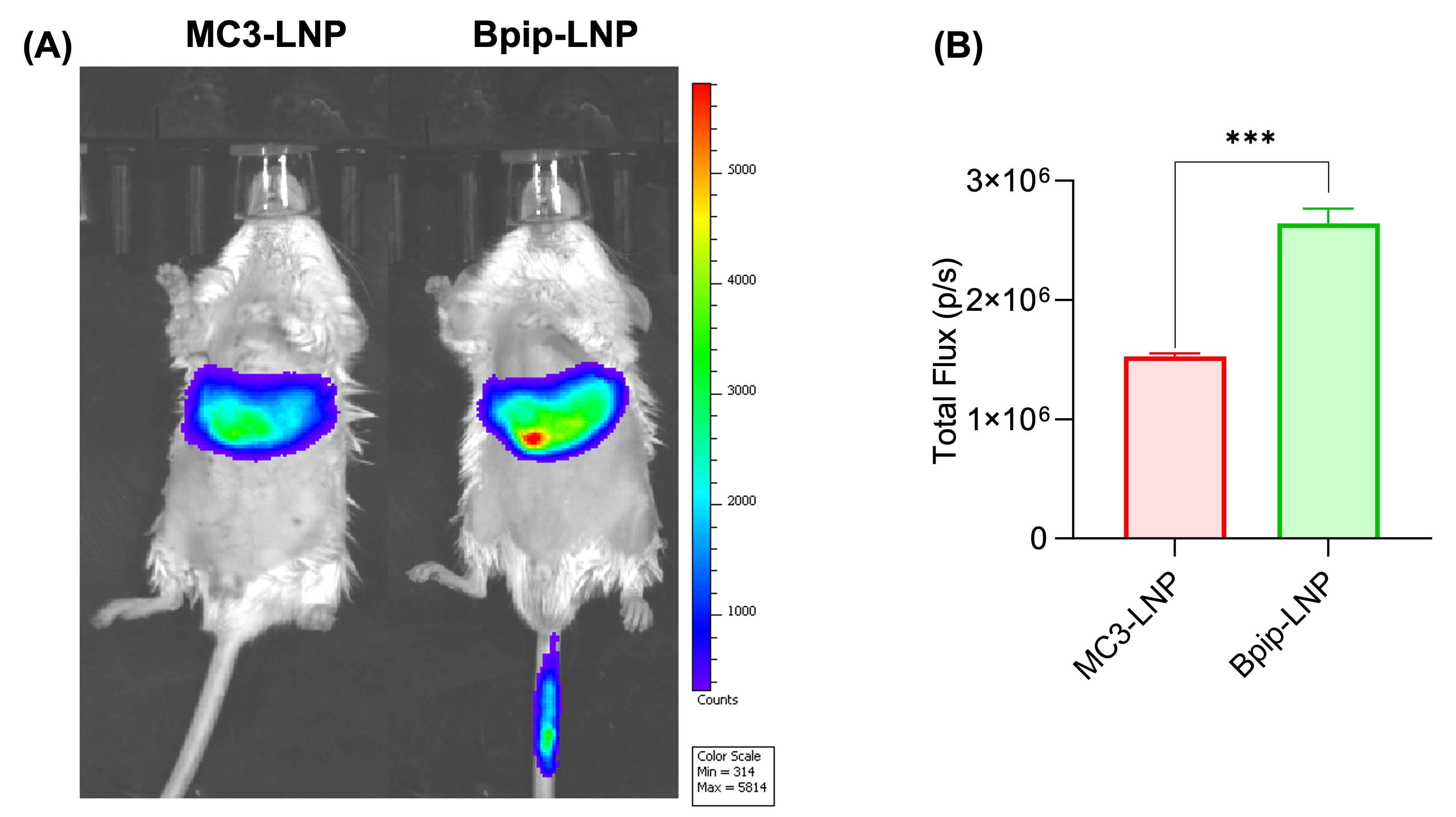 Figure 3. In vivo biodistribution of Bpip and MC3 LNPs. (A) A representative biodistribution image of MC3 and Bpip LNPs in BALB/c mice captured under IVIS. LNPs with a Fluc-mRNA dose of 0.5mg/kg were injected via the tail vein, and biodistribution was observed at 6h post-injection. (B) The luminescence intensity in the form of total flux in the MC3 and Bpip-treated mice was quantified. Bpip shows a significantly higher mRNA translation efficiency than MC3.