
Formulation and Delivery - Chemical
Solid-State Characterization of Crinone<sup>®</sup> (Progesterone Vaginal Gel): Microscopy, X-Ray Diffraction, and Thermal Behavior
 photo")
Ji Li, PhD (he/him/his)
Dr.
University of Michigan
Ann Arbor, Michigan, United States
Kaikai Wang, MS
Ph.D. candidate
University of Michigan
Ann Arbor, Michigan, United States
Yiran Huo, BS
Master Student
University of Michigan
Ann Arbor, Michigan, United States
Vivian Juang, MS
Ph.D. candidate
University of Michigan
Ann Arbor, Michigan, United States
Anna Schwendeman, Ph.D.
Larry and Ann Hsu Professor, Professor of Pharmaceutical Sciences
University of Michigan
Ann Arbor, Michigan, United States
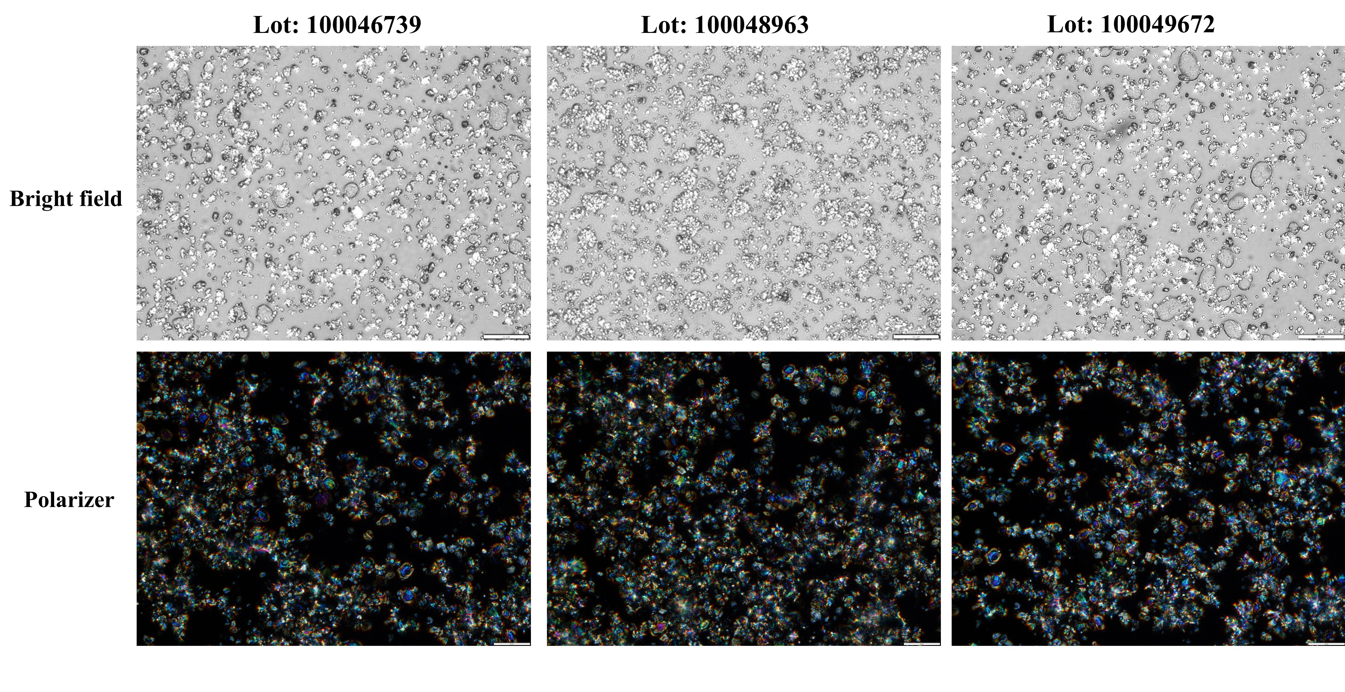 Figure 1. Microscopic images of three batches of Crinone® under bright field (upper panel) and polarized light (lower panel). The scale bar represents 20 μm.
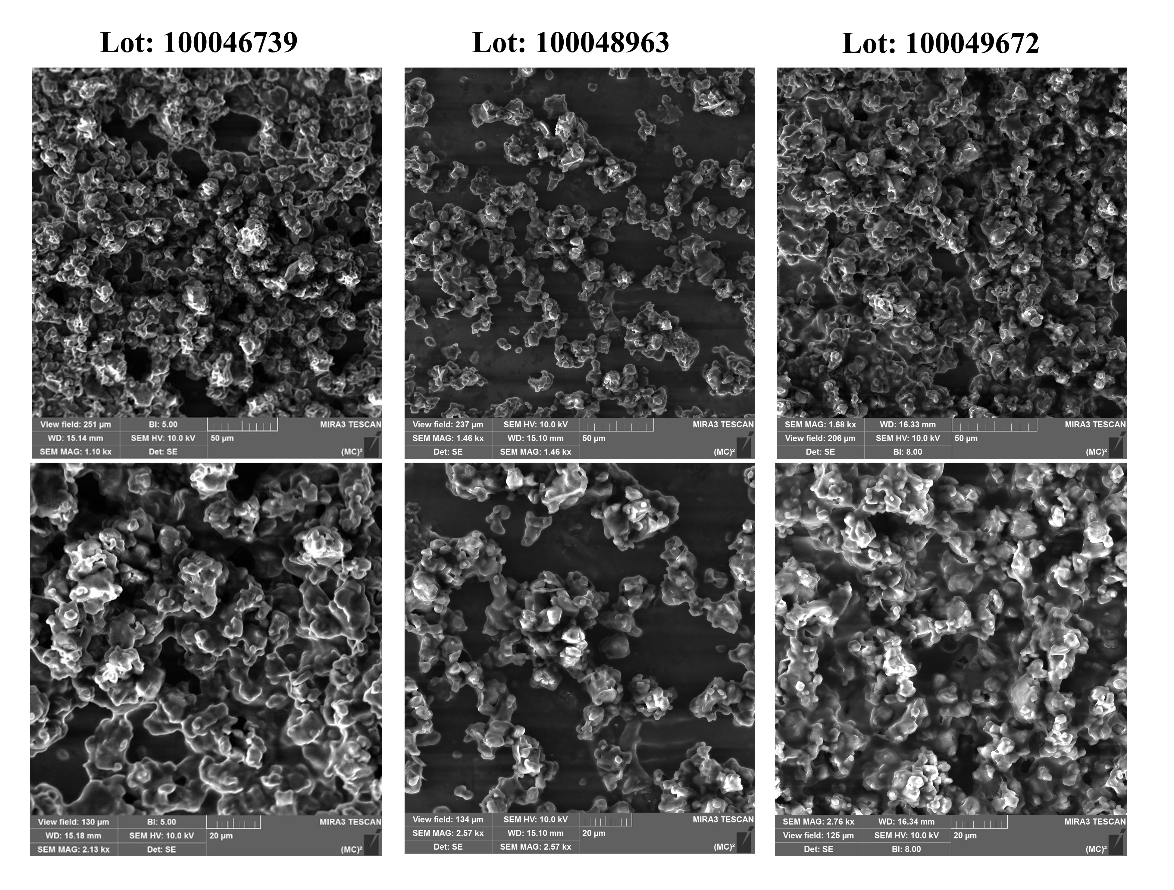 Figure 2. SEM images of three batches of Crinone®. The scale bar in the upper panel represents 50 μm and lower panel represents 20 μm.
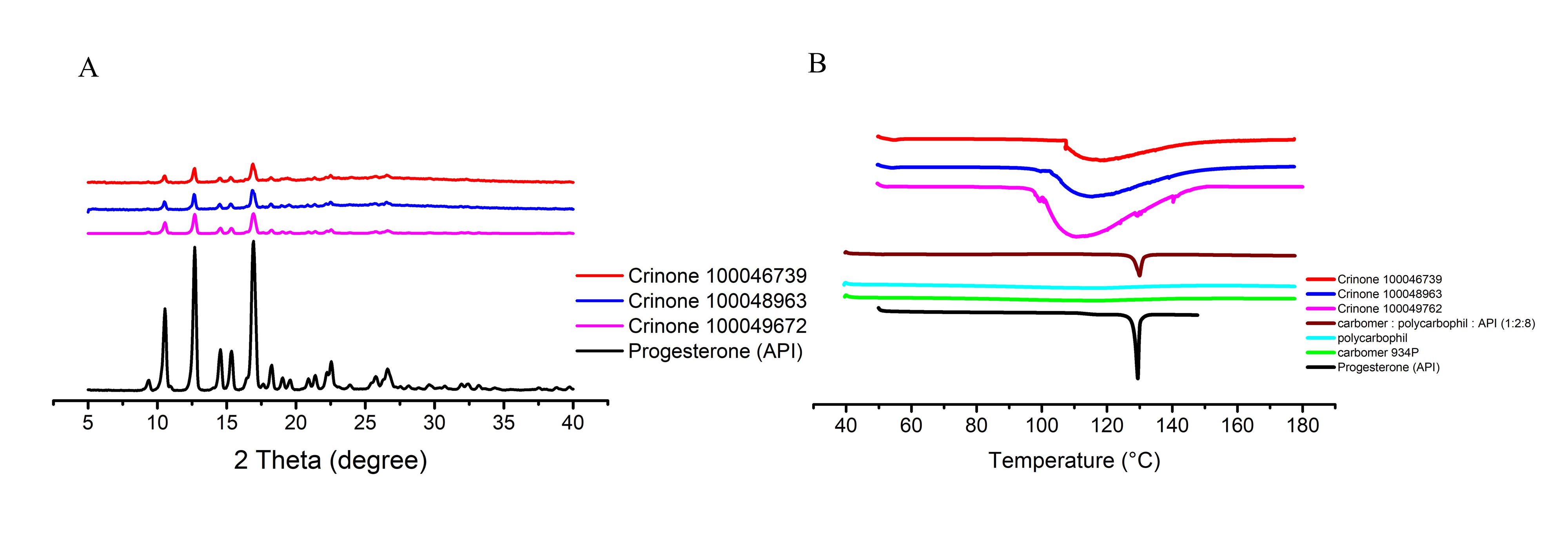 Figure 3. (A) X-rays diffraction patterns of progesterone (API) and three different batches of Crinone®. (B) Differential scanning calorimetry curves of different ingredients in Crinone® and formulations.