
Manufacturing and Analytical Characterization - Chemical
Shuaiqian Men, BS
Research Assistant
University of Maryland
Baltimore, Maryland, United States
Shuaiqian Men, BS
Research Assistant
University of Maryland
Baltimore, Maryland, United States

James E. Polli, Ph.D.
Professor and Ralph F. Shangraw/Noxell Endowed Chair in Industrial Pharmacy and Pharmaceutics
University of Maryland
Baltimore, Maryland, United States
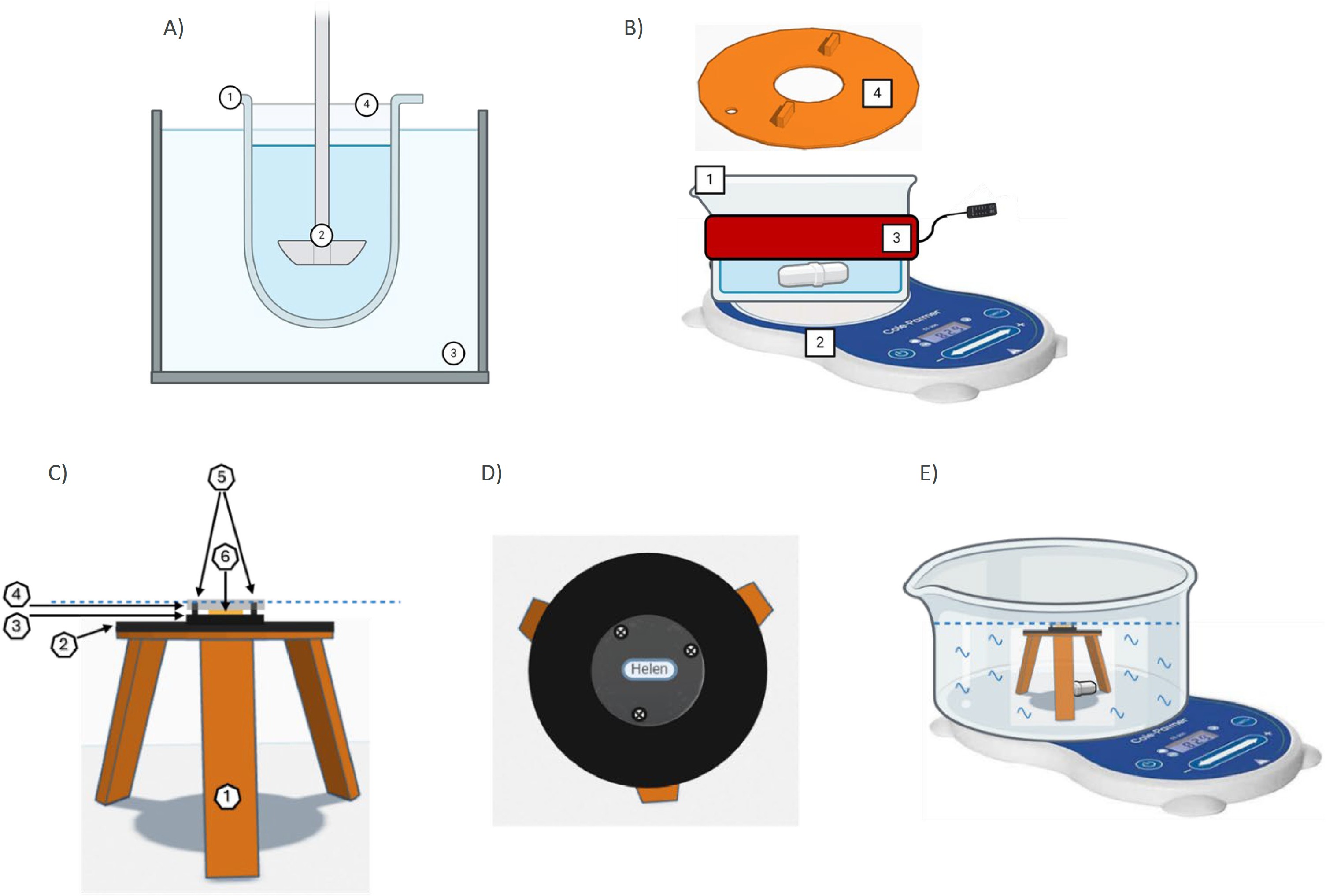 Fig. 1. Comparison of MeDDiS to USP II apparatus, and diagram of MeDDiS disc holder and ASD sandwich components. Panel A shows USP II apparatus. Panel B illustrates some MeDDiS components. Differences include: 1. MeDDiS employs a wide 1 L crystallization dish, allowing fit underneath microscope lens (not shown), in place of the compendial dissolution vessel. 2. MeDDiS uses a stir bar and stirring plate in place of a paddle. 3. MeDDiS employs a beaker heating wrap in place of a water heating bath. 4. MeDDiS uses a 3D printed lid (with hole for temperature monitoring) that allows microscope imaging (from above) of a centrally positioned ASD disc (not shown). Panel C is a side view of ASD disc holder, which consists of 1) 3D printed, 3-legged holder which sits in the dissolution vessel, 2) cuttable magnetic sheets, 3) 1-inch black acrylic plastic saucer below ASD disc (with name sticker to assess visual transparency of ASD disc), 4) 1-inch clear acrylic plastic saucer to protect ASD disc top from dissolution media, 5) mini-screws to compress to clear plastic saucer towards black acrylic plastic saucer and hence protect ASD disc top and bottom from dissolution media, and 6) ASD disc stabilized with silicone grease. The black acrylic saucer, the ASD disc, and the clear acrylic saucer are held together via the mini-screws and form the ASD sandwich. A magnetic sheet was fixed to the bottom of the black acrylic plastic saucer (i.e. the ASD sandwich) to stabilize it onto the center of the holder, via magnetic force. Panel D is a top view of ASD disc holder and ASD sandwich without an ASD disc. Name sticker “Helen” is visibly obscured when ASD disc is in a gel state. Panel E highlights the positioning of holder in the center of the dissolution vessel (secured with jelly tape) with stir bar on stirring plate. The dotted blue dotted line in panels A and C indicates the dissolution media level, which did not exceed the top clear acrylic plastic saucer, allowing the above microscope (not drawn) to focus on the dissolving ASD discs throughout the study.
.jpg) Fig. 2. Digital microscope images of four discs undergoing dissolution in MeDDiS. Four discs had compositions 20DL/80PL, 25DL/75PL, 30DL/70PL, and 40DL/60PL. A) 20DL/80PL was chosen as an example of low DL. B) 25DL/75DL showed slightly slower disc loss than discs with low DL. C) 30DL/70PL exhibited a large amount of remaining white gel-like undissolved particles. D) 40DL/60PL exemplified high DL where >80 % dry glassy core was undissolved at the end of the study at 300 min.
Fig. 2. Digital microscope images of four discs undergoing dissolution in MeDDiS. Four discs had compositions 20DL/80PL, 25DL/75PL, 30DL/70PL, and 40DL/60PL. A) 20DL/80PL was chosen as an example of low DL. B) 25DL/75DL showed slightly slower disc loss than discs with low DL. C) 30DL/70PL exhibited a large amount of remaining white gel-like undissolved particles. D) 40DL/60PL exemplified high DL where >80 % dry glassy core was undissolved at the end of the study at 300 min..jpg) Fig. 3. Comparison of observed and predicted RTV and PVPVA profiles from discs with different DLs/PLs. A) 5DL/95PL. B) 10DL/90PL. C) 15DL/85PL. D) 20DL/80DL. E) 25DL/75DL. F) 30DL/70PL. G) 35DL/65PL. H) 40DL/60PL. I) 45DL/55PL. J) 50DL/50PL. Observed profiles were measured using HPLC and SEC-UV for drug and polymer respectively. Predicted profiles were from microscope imaging. Error bar is SD from n = 3.
Fig. 3. Comparison of observed and predicted RTV and PVPVA profiles from discs with different DLs/PLs. A) 5DL/95PL. B) 10DL/90PL. C) 15DL/85PL. D) 20DL/80DL. E) 25DL/75DL. F) 30DL/70PL. G) 35DL/65PL. H) 40DL/60PL. I) 45DL/55PL. J) 50DL/50PL. Observed profiles were measured using HPLC and SEC-UV for drug and polymer respectively. Predicted profiles were from microscope imaging. Error bar is SD from n = 3.