
Formulation and Delivery - Chemical
Usha Y. Nayak, Ph.D.
Dr.
Manipal College of Pharmaceutical Sciences
Manipal, Karnataka, India
Ashwini T, M. Pharm
Dr.
Manipal College of Pharmaceutical Sciences
UDUPI, Karnataka, India
Hang T Nguyen, Ph.D.
Dr.
University of South Australia
Adelaide, South Australia, Australia
Raghu Chandrashekar H, Ph.D.
Dr.
Manipal College of Pharmaceutical Sciences
Udupi, Karnataka, India
Yogendra Nayak, PhD (he/him/his)
Dr.
Manipal College of Pharmaceutical Sciences
Udupi, Karnataka, India
Padmaja A A. Shenoy, M.D.
Dr.
Kasturba Medical College
Udupi, Karnataka, India
Abiodun D Ogunniyi, Ph.D.
Dr.
University of Adelaide
Adelaide, South Australia, Australia
Sanjay Garg, Ph.D.
Professor
University of South Australia
Adelaide, South Australia, Australia
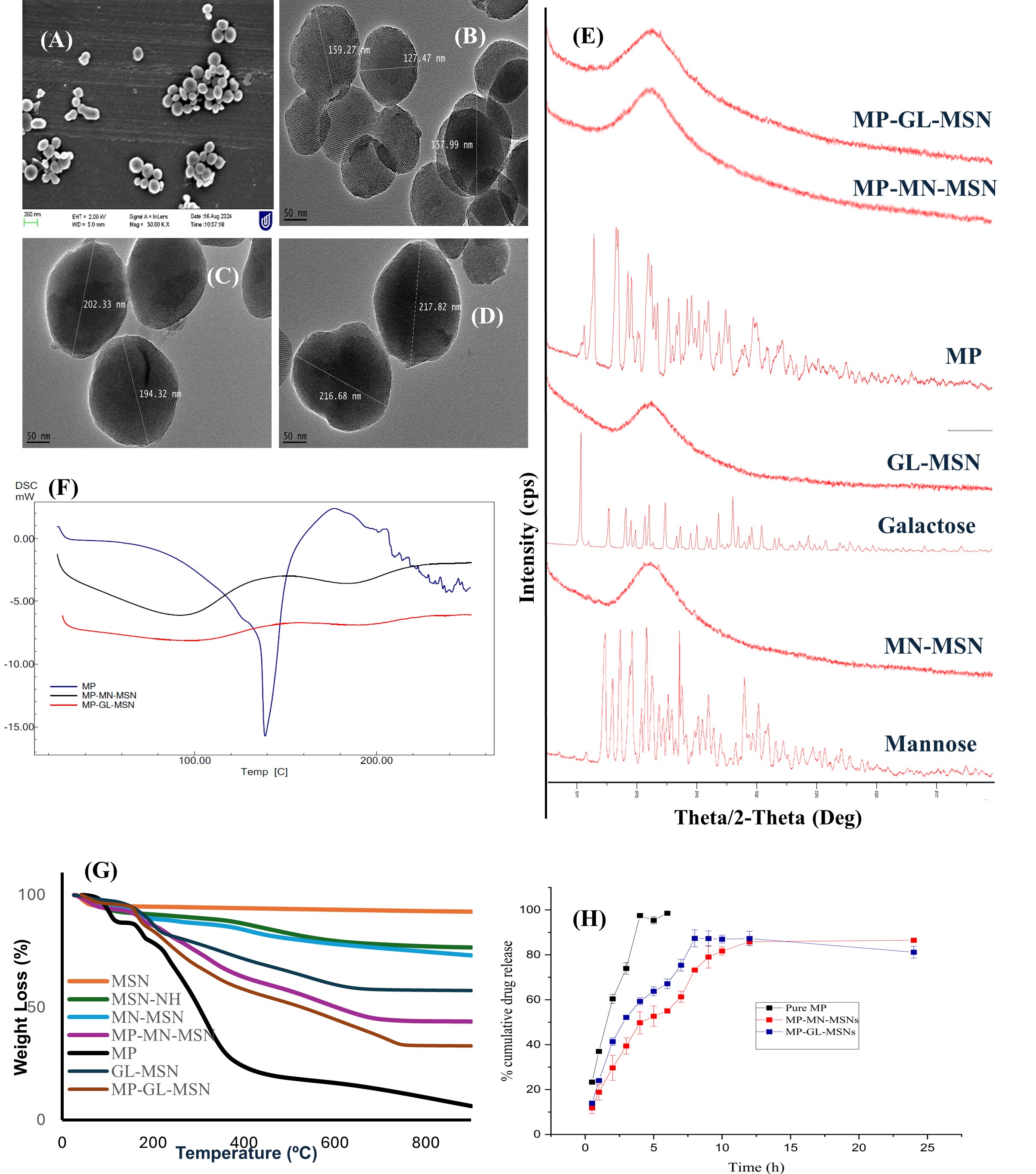 Fig.1. Characterization of MP loaded glyco-conjugated MSN. SEM images for (A) MSN-NH; TEM images for (B) GL-MSN (C) MP-MN-MSN (D) MP-GL-MSN; (E) XRD pattern for mannose, galactose, glyco-conjugated MSN, Pure MP, MP loaded Glyco-conjugated MSN; (F) DSC thermogram of Pure MP and MP loaded glyco-conjugated MSN; (G) TGA of MSN, amine functionalized MSN, glyco-conjugated MSN, Pure MP, MP loaded Glyco-conjugated MSN; (H) In vitro release profiles of meropenem from glyco-conjugated MSN.
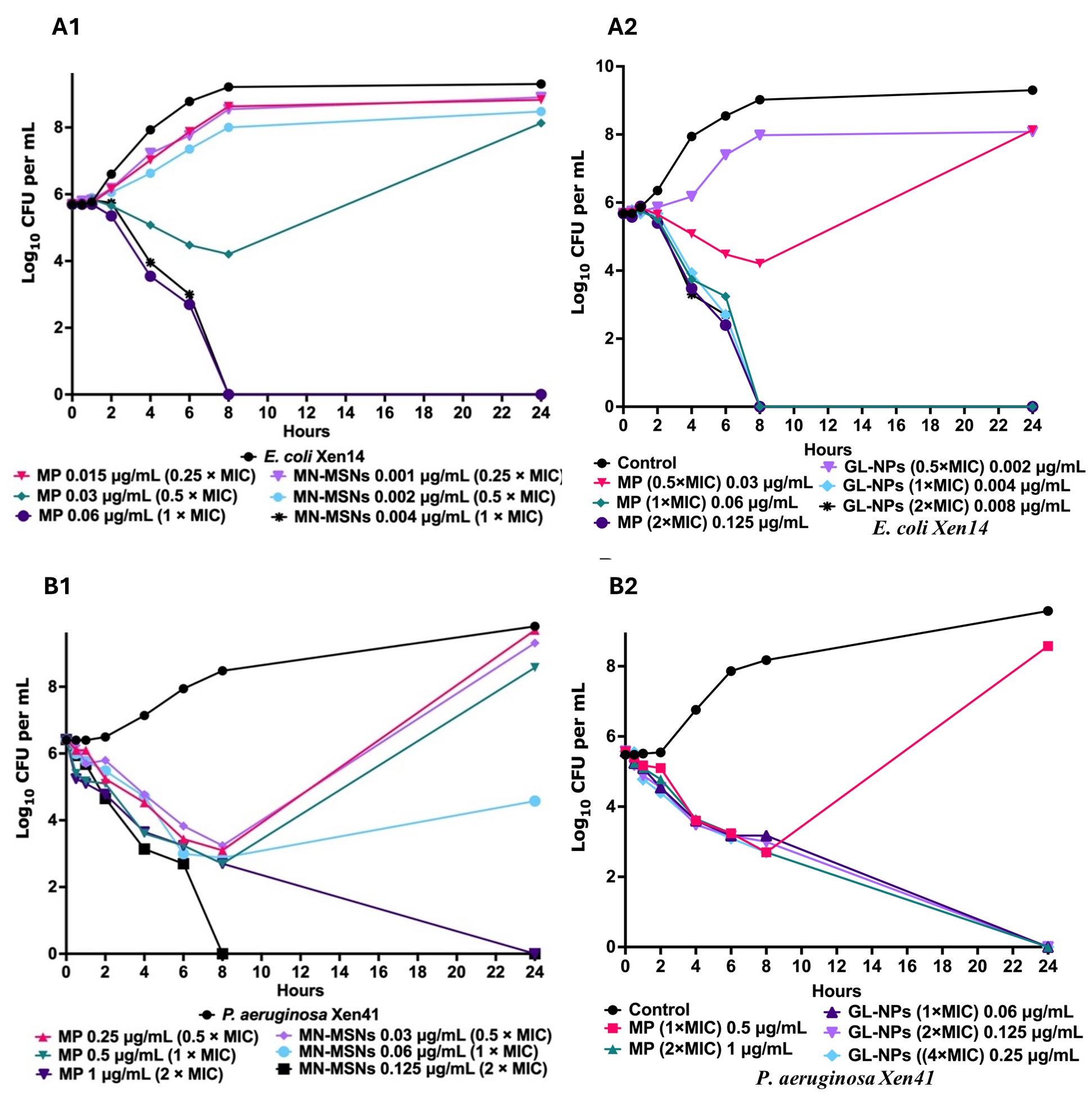 Fig. 2. Time- and concentration-killing and growth-inhibitory kinetics of MP, MP loaded MN-MSNs (MN-MSNs), and MP loaded GL-MSNs (GL-MSNs) against Gram-negative bacterial pathogens. MP, MN-MSNs and GL-MSNs were prepared at various concentration of 0.25 MIC, 0.5 MIC, 1 MIC, and 2 MIC. (A1, A2, B1, and B2) time-killing kinetics of MP loaded MN-MSNs and MP loaded GL-MSNs in comparison with MP against tested bacteria; tests were prepared in duplicate in 24 well-flat bottom plates (CLS3738, Sigma-Aldrich), then samples were withdrawn at indicated times for serial dilutions and plating and incubation at 37°C overnight on HBA plates. MIC, minimum inhibitory concentration.
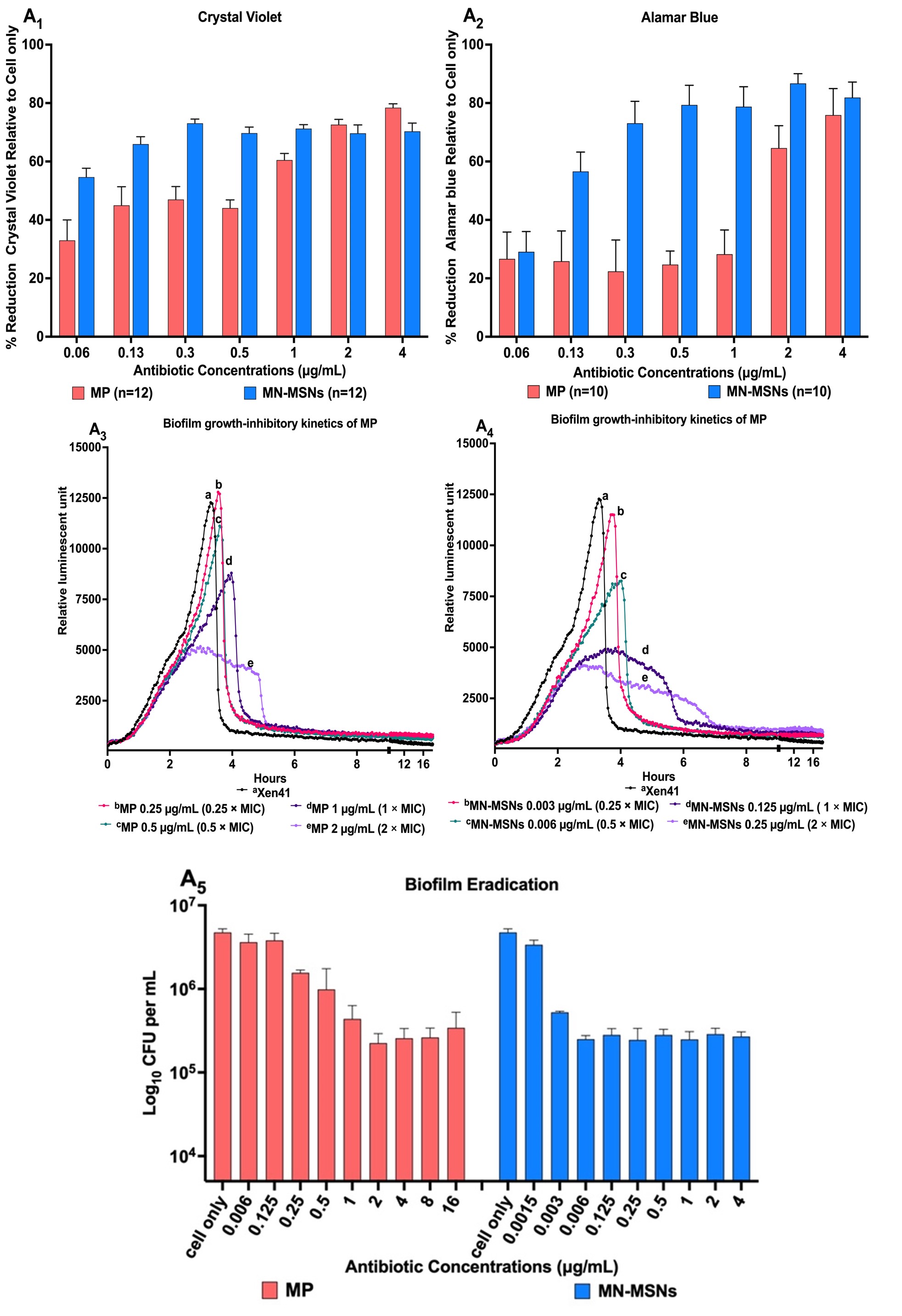 Fig.3. Comparison of anti-biofilm activities of MN-MSNs with MP against P. aeruginosa (Xen41). A mature biofilm of P. aeruginosa was generated by growth at 24 h in 96-well plates. (A1) presenting reduction in Xen41 biofilm mass following 24 h treatment with MP and MP-loaded MN-MSNs (MN-MSNs) was quantified using crystal violet staining. (A2) reduction in Xen41 viability in biofilm flowing 24 h treatment with MP and MN-MSNs, cell viability was quantified using Alamar Blue. (A3 & A4) showing time and concentration- Xen41 viability kinetics in biofilm measured by changes in bacterial metabolic activity relative to the reduction of Xen41 luminescence compared with growth control. (A5) presenting the total number of viable Xen41 cells remaining on pegs of MBEC lid after treatment with different concentrations of MN-MSNs and MP. Data represents mean standard deviation (n=12 [crystal violet]; n=10 [Alamar Blue]; n=4 [MBEC]).