
Formulation and Delivery - Biomolecular
Sara Aly Attia, BS
Ph.D. Candidate
University of Southern California
Los Angeles, California, United States
 photo")
John A. MacKay, PhD (he/him/his)
Professor
University of Southern California
LA, California, United States
Amanda Tse, BS
Ph.D. Student
University of Southern California
LA, California, United States
Ashley Ramirez, MS
Graduate student
University of Southern California
LA, California, United States
Paul Seidler, Ph.D.
Professor
University of Southern California
LA, California, United States
Ram Kannan, Ph.D.
Adjunct Professor & Senior Scientist, Doheny Eye Institute
University of California
LA, California, United States
Sreekumar Parameswaran, Ph.D.
Scientist, Doheny Eye Institute
University of California
LA, California, United States
John A. MacKay, PhD (he/him/his)
Professor
University of Southern California
LA, California, United States
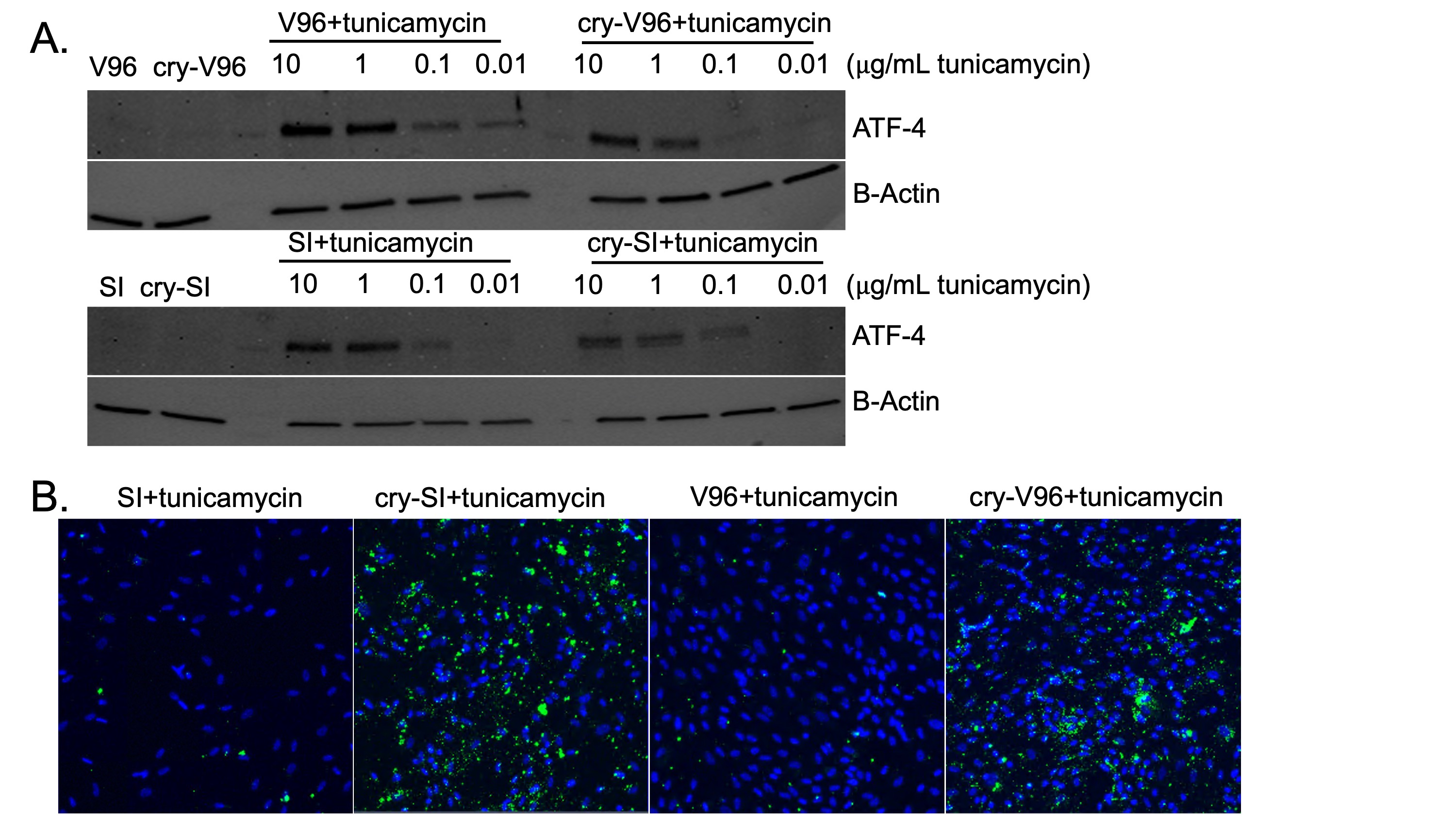 Figure 1. Cry-ELPs downregulate ATF-4 and enhance cellular association under tunicamycin-induced ER stress. (A) WB was used to evaluate the effects of pre-treating ARPE-19 cells with 25 μM of ELP-based formulations for 24 h, followed by a 24 h challenge with a tunicamycin gradient (10-0.01 μg/mL). As shown, only cry-SI and cry-V96 significantly reduced ATF-4 levels, compared to their respective controls. B) Microscopy data further confirmed that only cry-ELPs exhibited enhanced cellular association under tunicamycin-induced stress.
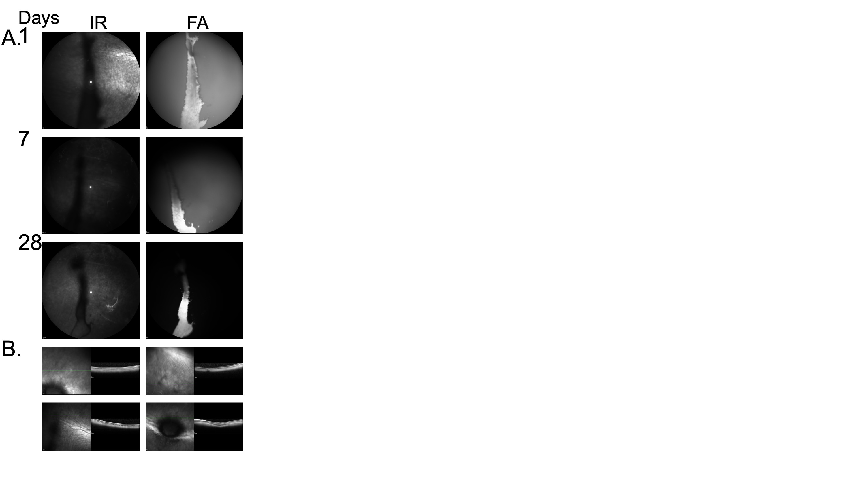 Figure 2. Cry-V96 demonstrates prolonged intravitreal retention over 28 days following a single intravitreal injection, while maintaining retinal integrity. A) Spectralis OCT was used to monitor the pharmacokinetics of a single intravitreal injection of ~ 300 μM fluorescein-labeled cry-V96 in NZP rabbits. Infrared reflectance (IR) and fluorescein angiography (FA) imaging revealed a visible depot throughout the 28-day period. Diffusion from the depot (indicated by whitish background signal) was clearly observed on days 1 and 7. B) Retinal sections obtained on day 28 at different spots, prior to euthanasia, showed preserved retinal structure, confirming the absence of observable toxicity.