
Preclinical, Clinical, and Translational Sciences

Huanhuan Li, Ph.D.
Research Fellow
Queen's University Belfast
BELFAST, Northern Ireland, United Kingdom
Huanhuan Li, Ph.D.
Research Fellow
Queen's University Belfast
BELFAST, Northern Ireland, United Kingdom
 photo")
Ryan Donnelly, Ph.D. (he/him/his)
Professor
Queen's University Belfast
Belfast, Northern Ireland, United Kingdom
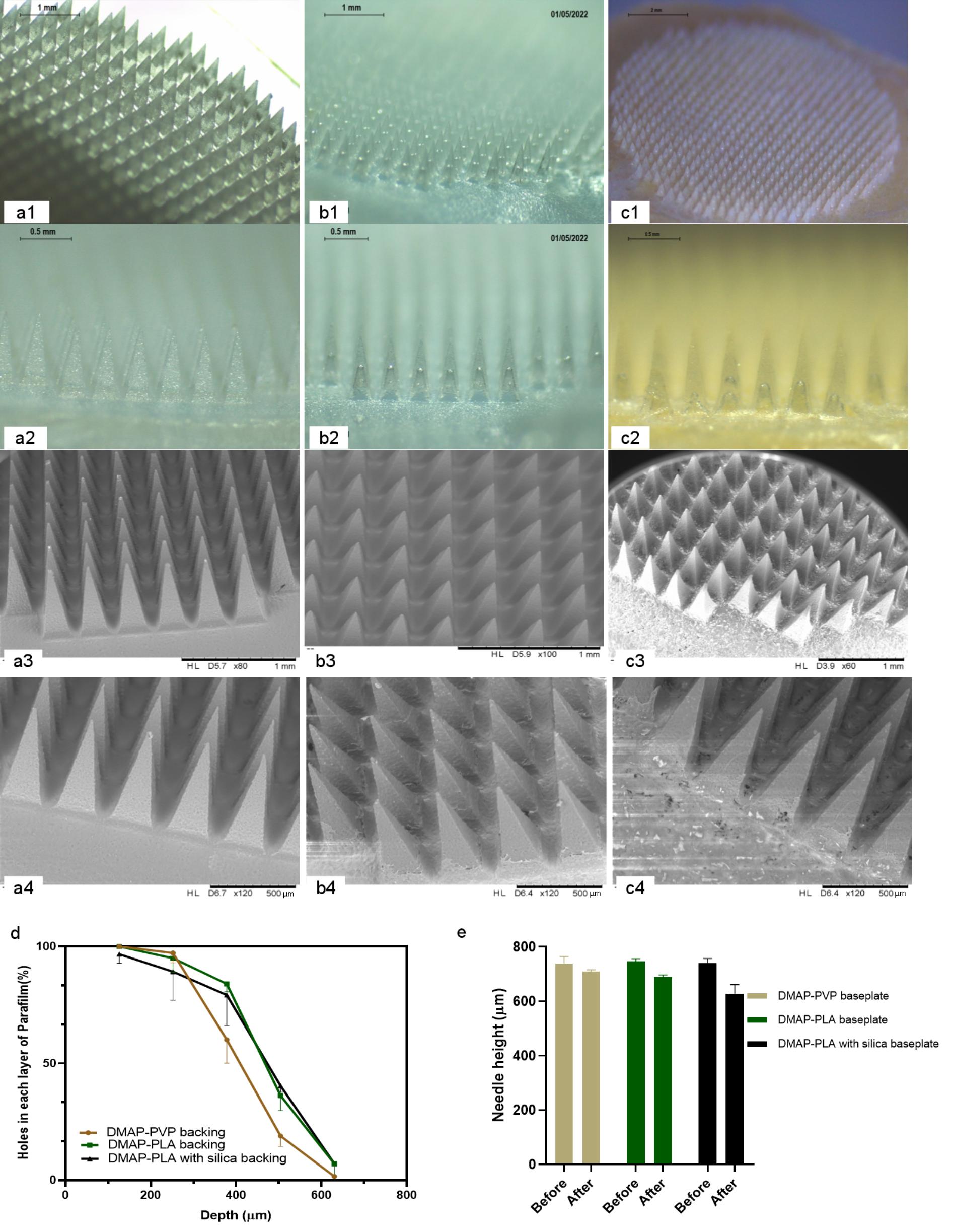 Figure 1. Morphology of PVP-DMAPs (a1-4), PLA-DMAPs (b1-4) and PLA silica-DMAPs (c1-4) obtained by optical microscope and SEM. Results of the Parafilm®M insertion test (d) (Means-SDs, n=5) and height reduction test (Means + SDs, n=5) (e) of DMAPs with PVP, PLA and PLA with a silica infued-baseplate.
 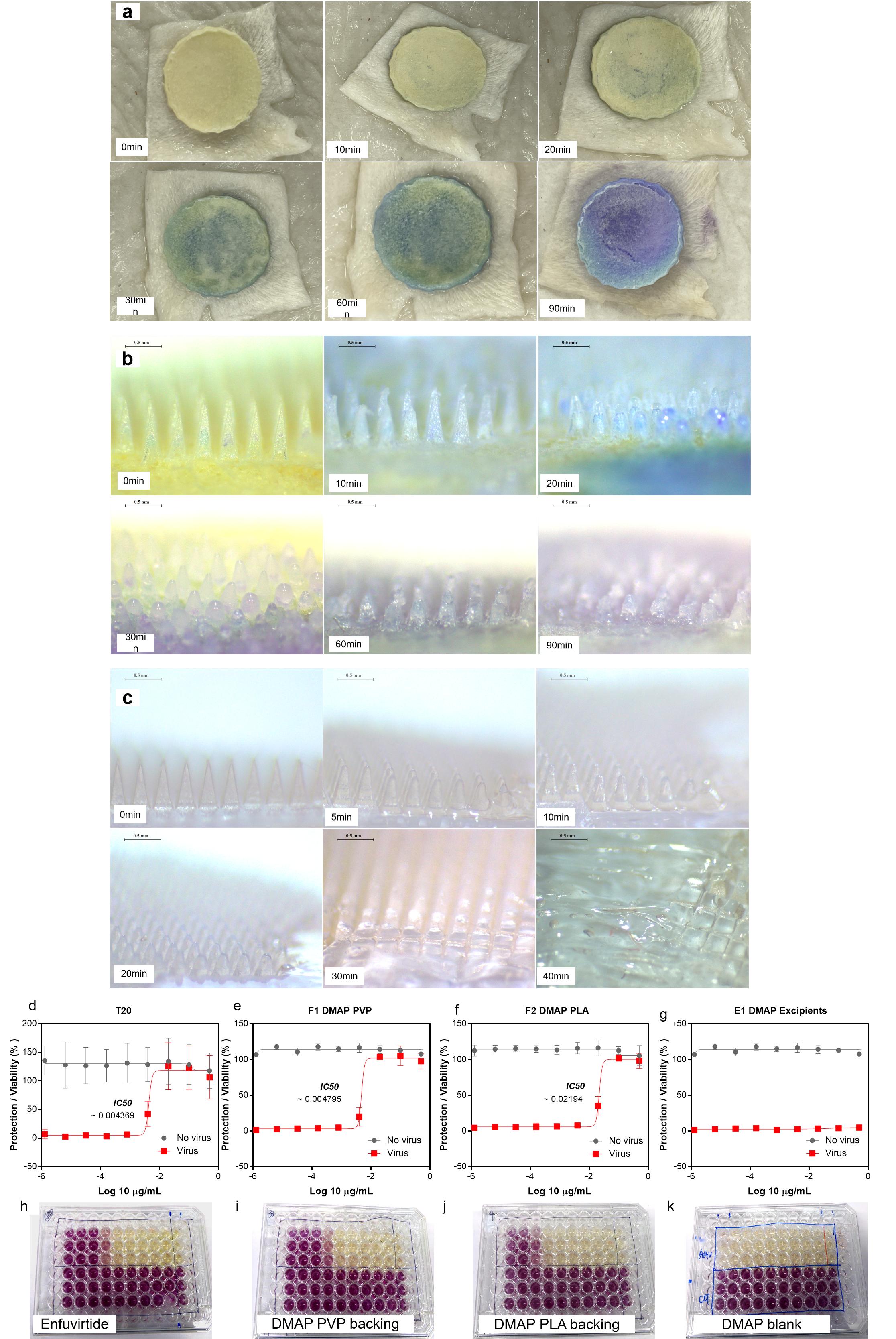 Figure 2. Schematic illustration of the fabrication process of DMAPs (a0). Morphology of dissolving microneedles of FITC-dextran (a-f) and fluorescein sodium (g-l) before (a-c&g-i) and after (d-f&j-l) needle dissolution obtained by optical microscopy and SEM. Representative fluorescence images and Z-stack images of DMAP of FITC-dextran 150 kDa (m, n, o) and fluorescein sodium (p, q, r) obtained by fluorescence microscope and multiphoton microscopy. The percentage of viable cells after 72 h of culture with PLA+M180 and PLA+M50 DMAPs in MTT assay (Means + SD, n=6) (k). Alive/dead staining of fibroblastic cells on control, PLA+M180 and PLA+M50 treated samples (l) (Green represented alive and red represented dead). Total DNA content of cells on control, PLA+M180 and PLA+M50 cultured for 72 h in PicoGreen assay (Means + SD, n=6) (m). ** represents p<0.001.
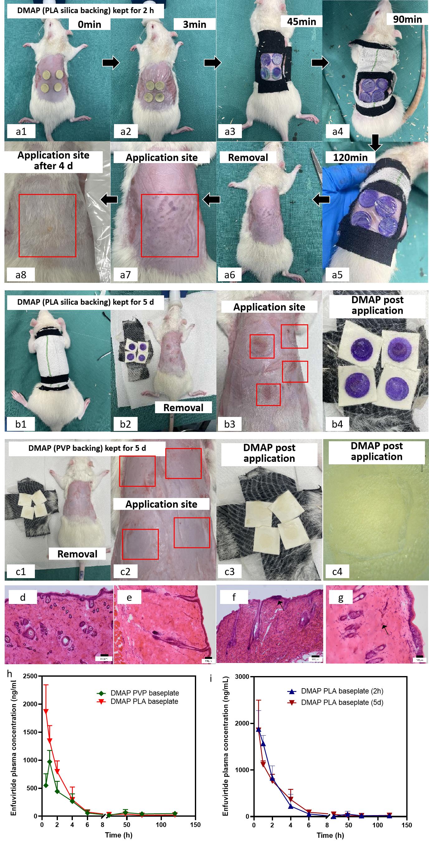 Figure 3. Ex vivo dissolution (a, c) and baseplate colour change (b,d) for FITC-dextran (a, b) and fluorescein sodium (c, d) dissolving microneedle. Ex vivo delivery of the two dissolving microneedles (e) and distribution of the analytes (mean±SD, n = 3) (f). Preliminary result from dissolving microneedles with the feedback system on rats (g).