
Discovery and Basic Research
Manali Patel, MS
PhD Candidate
University at Buffalo
Buffalo, New York, United States
Manali Patel, MS
PhD Candidate
University at Buffalo
Buffalo, New York, United States
 photo")
Vincent Chak, PhD (he/him/his)
PhD Candidate
University at Buffalo
Buffalo, New York, United States
Beverly Schaefer, M.D.
Medical Director
WNYBloodCare (formerly Hemophilia Center of WNY)
buffalo, New York, United States

Sathy V. Balu-Iyer, Ph.D.
Professor
University at Buffalo
Buffalo, New York, United States
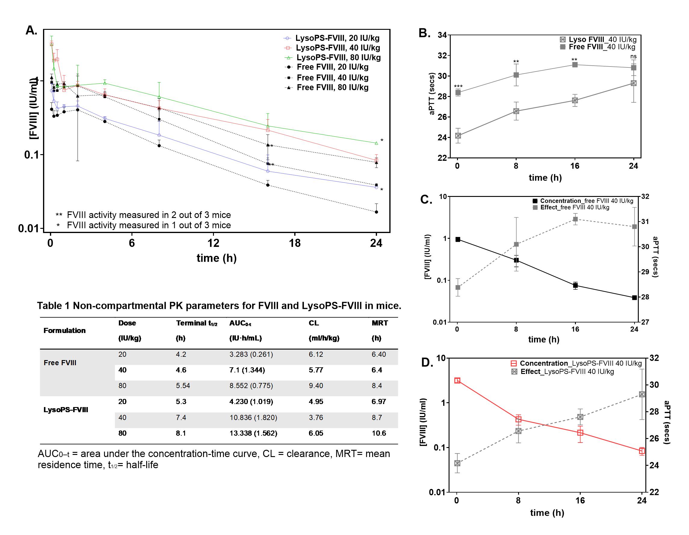 Fig 1: Pharmacokinetics profiles of FVIII and LysoPS-FVIII in HA mice. A) Plasma concentration-time profiles following IV administration of free FVIII at 20 (closed circles), 40 (closed square), or 80 (closed triangle) IU/kg or LysoPS-FVIII at 20 (open circles), 40 (open square), or 80 (open triangle) IU/kg. Table 1: Pharmacokinetic parameters determined by non-compartmental analysis. B) Ex vivo hemostatic efficacy measured using activated partial thromboplastin time (aPTT) assay in HA mice at dose 40IU/kg. Statistics were performed using two-way ANOVA-Bonferroni’s multiple comparison test (*P < 0.05). Overlay of PK and PD profiles after C) free FVIII treatment D) after LysoPS-FVIII.
.jpg) Fig 2: Simulated clinical PK profiles and projected impact of LysoPS association on prophylactic free FVIII administration. A) Model fitting for LysoPS-FVIII using a 2-compartment linear clearance model (solid lines). Model fitted parameters for LysoPS-FVIII (Table 2). B) Projected human PK profile of LysoPS-FVIII (red) and observed disposition of free FVIII in humans (black) at 40 IU/kg doses. Literature-derived parameters were used for free FVIII simulations, while scaled human PK parameters were predicted using an “informed scaling” approach combining allometric PK parameter scaling with normalized Wajima curves (Table 3).
Fig 2: Simulated clinical PK profiles and projected impact of LysoPS association on prophylactic free FVIII administration. A) Model fitting for LysoPS-FVIII using a 2-compartment linear clearance model (solid lines). Model fitted parameters for LysoPS-FVIII (Table 2). B) Projected human PK profile of LysoPS-FVIII (red) and observed disposition of free FVIII in humans (black) at 40 IU/kg doses. Literature-derived parameters were used for free FVIII simulations, while scaled human PK parameters were predicted using an “informed scaling” approach combining allometric PK parameter scaling with normalized Wajima curves (Table 3)..jpg) Figure 3: Biodistribution studies A) Whole-body imaging post retro orbital injection of either free indocyanine green (ICG) or ICG-loaded LysoPS nanoparticles in Swiss Webster mice. B) Organs were collected at 1- and 3-hours post-injection and imaged using the FMT 2000 In Vivo Imaging System. Liver localization was further confirmed using confocal microscopy analysis post retro-orbital injection of C) buffer D) Dil dye-labeled LysoPS nanoparticles (DAPI-Blue, Liposome- Yellow, LYVE1- Green) E) Comparison of Mean fluorescence intensity (MFI).
Figure 3: Biodistribution studies A) Whole-body imaging post retro orbital injection of either free indocyanine green (ICG) or ICG-loaded LysoPS nanoparticles in Swiss Webster mice. B) Organs were collected at 1- and 3-hours post-injection and imaged using the FMT 2000 In Vivo Imaging System. Liver localization was further confirmed using confocal microscopy analysis post retro-orbital injection of C) buffer D) Dil dye-labeled LysoPS nanoparticles (DAPI-Blue, Liposome- Yellow, LYVE1- Green) E) Comparison of Mean fluorescence intensity (MFI).